3D bioprinted ear cartilage nears clinical trials. Explore the technology, benefits, and future of bio medical 3d printing. Discover how LAVA3DP supports this innovation. Contact us for advanced 3D printing solutions.
For decades, reconstructive surgery for patients with congenital ear deformities, such as microtia, or those who have suffered traumatic injuries has relied on a challenging combination of autologous cartilage grafts or synthetic implants. These traditional methods often involve complex surgeries, donor site morbidity, and results that may not perfectly match the natural architecture of the ear. Today, a new frontier is emerging: 3D bioprinting of living ear cartilage. This technology is rapidly advancing from a promising concept to a tangible clinical reality, poised to revolutionize craniofacial reconstruction.
This article explores the science, recent breakthroughs, and future trajectory of 3D printed ear cartilage, examining the clinical, regulatory, and technical milestones that are bringing this innovation to patients. As a leader in advanced manufacturing, LAVA3DP provides the high-precision 3D printing equipment and expertise that underpin such transformative biomedical applications.
The Clinical Need: Why the Ear is a Prime Target for Bioprinting
The human ear’s intricate, three-dimensional structure makes it one of the most challenging anatomical features to reconstruct. Microtia, a congenital condition where the external ear is underdeveloped, occurs in approximately 1 in every 4,000 to 10,000 births globally. Traditional reconstruction, using rib cartilage harvested from the patient, is a demanding procedure requiring significant surgical skill. It involves two to four surgeries, extended hospital stays, and carries risks of chest wall deformity and scarring.
Synthetic implants, such as porous polyethylene, offer an alternative but lack the flexibility of native cartilage and carry a risk of extrusion or infection over time. This unmet clinical need has driven researchers toward tissue engineering, with the goal of creating a living, patient-specific ear construct that matures into natural tissue.
| Reconstruction Method | Key Characteristics | Limitations |
|---|---|---|
| Autologous Rib Cartilage Graft | Uses patient’s own tissue; gold standard for biocompatibility. | Invasive harvest; requires high surgical skill; donor site morbidity; limited by cartilage availability. |
| Synthetic Implants (e.g., Porous Polyethylene) | Readily available; no donor site. | No growth potential; risk of infection, extrusion, and material fatigue over time. |
| 3D Bioprinted Cartilage | Patient-specific geometry; uses living cells; potential for natural tissue integration. | Technology still under clinical validation; complex regulatory pathway; cost. |
The Science Behind 3D Bioprinted Cartilage
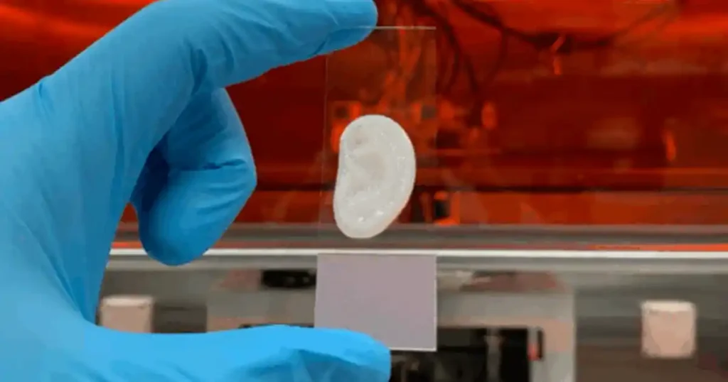
Creating a functional ear cartilage construct requires a convergence of several advanced technologies. The process typically begins with a 3D scan of a patient’s healthy ear (or a mirrored image of the contralateral ear) to create a digital blueprint. This blueprint guides a 3D bioprinter, which precisely deposits layers of a bio-ink.
A bio-ink is a carefully formulated mixture of living cells—usually chondrocytes (cartilage cells) derived from the patient or a donor, or mesenchymal stem cells (MSCs)—and a hydrogel scaffold material, such as alginate, gelatin, or decellularized extracellular matrix. The scaffold provides a temporary structure for the cells to adhere, proliferate, and produce their own extracellular matrix. After printing, the construct is cultured in a bioreactor that mimics the physiological environment, allowing the cells to mature into robust, flexible cartilage tissue.
Key Advantages of Bioprinting for Ear Cartilage:
- Patient-Specific Anatomy: The construct is designed to precisely match the patient’s unique anatomy, improving aesthetic outcomes.
- Reduced Surgical Morbidity: Eliminates the need for rib cartilage harvest, simplifying the surgical procedure.
- Living Tissue: The resulting construct is composed of the patient’s own cells (in autologous approaches), minimizing the risk of immune rejection and integrating as natural tissue.
- Scalability: The process is digitized, potentially allowing for greater reproducibility compared to hand-carved grafts.
Recent Milestones: Moving Toward Clinical Reality
While the concept has been explored for over a decade, the past few years have witnessed a significant acceleration toward clinical application. Several research groups and companies have reported promising results in both preclinical animal models and first-in-human trials.
1. Preclinical Success in Large Animal Models
Long-term studies in sheep and swine models have been instrumental. For instance, a 2023 study published in Cell Reports Medicine demonstrated that 3D bioprinted ear cartilage constructs, when implanted in sheep, maintained their shape and developed mature cartilage tissue with biomechanical properties comparable to native ear cartilage over a 12-month period. This study was pivotal in demonstrating the long-term stability and safety of the constructs.
2. First-in-Human Clinical Trials
The most significant leap forward came with the initiation of the first prospective, multicenter clinical trials evaluating 3D bioprinted ear cartilage for microtia reconstruction. Companies like 3DBio Therapeutics (now part of a larger entity) initiated a pivotal trial where a 3D-bioprinted, patient-specific ear implant was surgically placed. Early results, presented at medical conferences, have shown encouraging outcomes with a positive safety profile and high patient satisfaction regarding the aesthetic result.
| Trial Parameter | Details |
|---|---|
| Design | Prospective, open-label, single-arm, multicenter trial. |
| Population | Patients with unilateral microtia, aged 5 years and older. |
| Intervention | Autologous, patient-specific 3D-bioprinted ear cartilage implant. |
| Primary Endpoint | Safety (adverse events, implant integrity) and aesthetic outcome at 12 months. |
| Status | Active, with published early data showing successful engraftment and cartilage formation. |
3. Regulatory Pathways
The U.S. Food and Drug Administration (FDA) has recognized the potential of 3D bioprinted tissues. These products are typically regulated as combination products (a medical device plus a biological component). The FDA has granted several designations, such as Orphan Drug Designation and Rare Pediatric Disease Designation, to expedite the development of treatments for microtia. This regulatory support has been crucial in de-risking the pathway to market for developers.
Challenges and Considerations for Widespread Adoption
Despite the promise, several hurdles remain before 3D bioprinted ear cartilage becomes a standard-of-care procedure.
1. Regulatory and Manufacturing Complexity
Scaling from a bespoke, laboratory-based process to a standardized, commercial manufacturing process is a formidable challenge. Each implant is patient-specific, requiring a GMP-compliant (Good Manufacturing Practice) workflow for cell isolation, expansion, bio-ink formulation, and printing. Maintaining sterility, viability, and quality control for each unique construct is a complex endeavor.
2. Long-Term Durability and Integration
While short- and mid-term data is promising, the long-term performance of bioprinted cartilage in the human body over decades is unknown. Questions remain about how the tissue will age, respond to mechanical stress, and maintain its shape and biomechanical properties over a patient’s lifetime.
3. Cost and Reimbursement
The cost of a personalized, cell-based therapy is currently substantial. Successful market adoption will depend on securing clear reimbursement pathways from healthcare payers. The technology must demonstrate not only superior clinical outcomes but also cost-effectiveness compared to traditional multi-stage surgeries.
The Role of Advanced 3D Printing Equipment
The realization of clinical-grade 3D bioprinted ear cartilage would not be possible without the underlying hardware and software that enable extreme precision and reproducibility. This is where companies like LAVA3DP play a vital role. The equipment used in these applications goes far beyond conventional fused deposition modeling.
LAVA3DP specializes in advanced 3D printing technologies that are critical for biomanufacturing, including:
- High-Resolution Stereolithography (SLA) and Digital Light Processing (DLP): Used to create the precise, biocompatible molds and temporary scaffolds required for tissue engineering.
- Micro-Extrusion Bioprinters: Systems capable of handling viscous bio-inks containing living cells with micron-level precision to replicate the complex architecture of auricular cartilage.
- Rigorous Quality Control: The need for repeatable, validated manufacturing processes aligns with LAVA3DP’s expertise in providing industrial-grade 3D printing solutions that meet strict quality management standards.
By providing the foundational technology for precision manufacturing, LAVA3DP empowers researchers and medical device manufacturers to translate laboratory innovations into clinically viable products.
| 3D Printing Technology | Application in Ear Cartilage Bioprinting |
|---|---|
| High-Resolution SLA/DLP | Printing of sacrificial molds to define the ear shape; fabrication of custom bioreactor chambers. |
| Micro-Extrusion Bioprinting | Direct deposition of cell-laden hydrogels to construct the living cartilage tissue layer by layer. |
| Laser-Assisted Bioprinting | High-precision placement of cells at high densities for research into complex tissue architecture. |
| Industrial 3D Printers | Scalable, GMP-compliant manufacturing of non-biological components of the implant system. |
The Future Outlook
The trajectory of 3D printed ear cartilage is a bellwether for the broader field of bio medical 3d printing. The coming years will be critical as ongoing clinical trials conclude and companies submit applications for regulatory approval. Several key developments will shape the future:
- Expansion of Applications: The success in ear reconstruction will pave the way for other craniofacial applications, including nasal reconstruction, orbital floor repair, and mandibular implants.
- Allogeneic (Off-the-Shelf) Products: To reduce costs and lead times, companies are exploring allogeneic cell sources—using donor cells that are universally compatible—to create “off-the-shelf” constructs that do not require a lengthy patient-specific cell culture phase.
- Integrated Bioprinting in Surgery: Advances in portable bioprinting and intraoperative imaging could one day allow surgeons to print and implant customized tissue constructs in a single surgical procedure.
- Synergy with AI and Automation: Machine learning algorithms will optimize bio-ink formulations and printing parameters, while automated, closed-loop bioprinting systems will ensure consistent, high-quality production.
Conclusion
The journey of 3D printed ear cartilage from a visionary concept to the threshold of clinical reality exemplifies the transformative potential of additive manufacturing in medicine. By combining patient-specific design, living cells, and precision engineering, this technology offers a profound improvement over traditional reconstructive methods. While challenges related to regulation, manufacturing scale-up, and cost persist, the robust clinical data and dedicated regulatory pathways signal a clear path forward.
For institutions and companies engaged in this cutting-edge field, access to reliable, high-performance 3D printing infrastructure is non-negotiable. LAVA3DP is committed to supporting the biomedical community with the advanced equipment and expertise required to turn these life-changing innovations into accessible clinical solutions. To learn how our technologies can empower your next project in bio medical 3d printing, please contact our team.
Frequent Asked Questions (FAQs)
1. What is bio medical 3d printing, and how does it differ from standard 3D printing?
Bio medical 3d printing encompasses the use of additive manufacturing to create medical devices, implants, and living tissues. Unlike standard 3D printing which uses plastics or metals, biomedical applications often require biocompatible or bioresorbable materials. In bioprinting, living cells are used to construct functional tissue. LAVA3DP provides the high-precision printing systems needed for both non-biological medical devices and the supportive structures for bioprinting.
2. Is 3D bioprinted cartilage already available for patients?
3D bioprinted ear cartilage is currently being evaluated in clinical trials and is not yet a broadly available commercial treatment. Early results are promising, with regulatory bodies like the FDA facilitating expedited review pathways. Access is currently limited to patients enrolled in these trials. LAVA3DP supports the developers and research institutions conducting these trials with advanced manufacturing equipment.
3. What materials are used in 3D printing for medical implants?
The materials vary widely depending on the application. For non-biological implants, biocompatible polymers like PEEK (polyetheretherketone) and titanium are common. For 3D bioprinting of tissues like cartilage, materials include hydrogels (such as alginate or gelatin) combined with a patient’s own cells. LAVA3DP’s equipment is compatible with a broad range of high-performance materials used in medical device manufacturing.
4. How does LAVA3DP ensure the precision required for medical 3D printing?
LAVA3DP specializes in industrial-grade 3D printing systems known for their high resolution and repeatability. Our technologies, including advanced SLA and DLP systems, achieve micron-level accuracy, which is essential for creating patient-specific anatomical models, surgical guides, and the intricate scaffolds used in tissue engineering. We provide comprehensive support to help clients meet stringent regulatory and quality standards.
5. How can I start a bio medical 3d printing project with LAVA3DP?
To begin a project, you should first define your application—whether it’s for surgical planning, a permanent implant, or a bioprinted tissue scaffold. LAVA3DP offers a range of systems and technical consultation to match your specific requirements. Our team can assist with material selection, hardware configuration, and process validation. Please contact us to discuss your project in detail.